xvr: system matches intraoperative X-rays to a 3D scan in seconds
Researchers at MIT and collaborating institutions described in Nature a system that lines up a two-dimensional X-ray image with a preoperative CT or MRI to sub-millimeter accuracy.
 Sofia MarchettiScience and health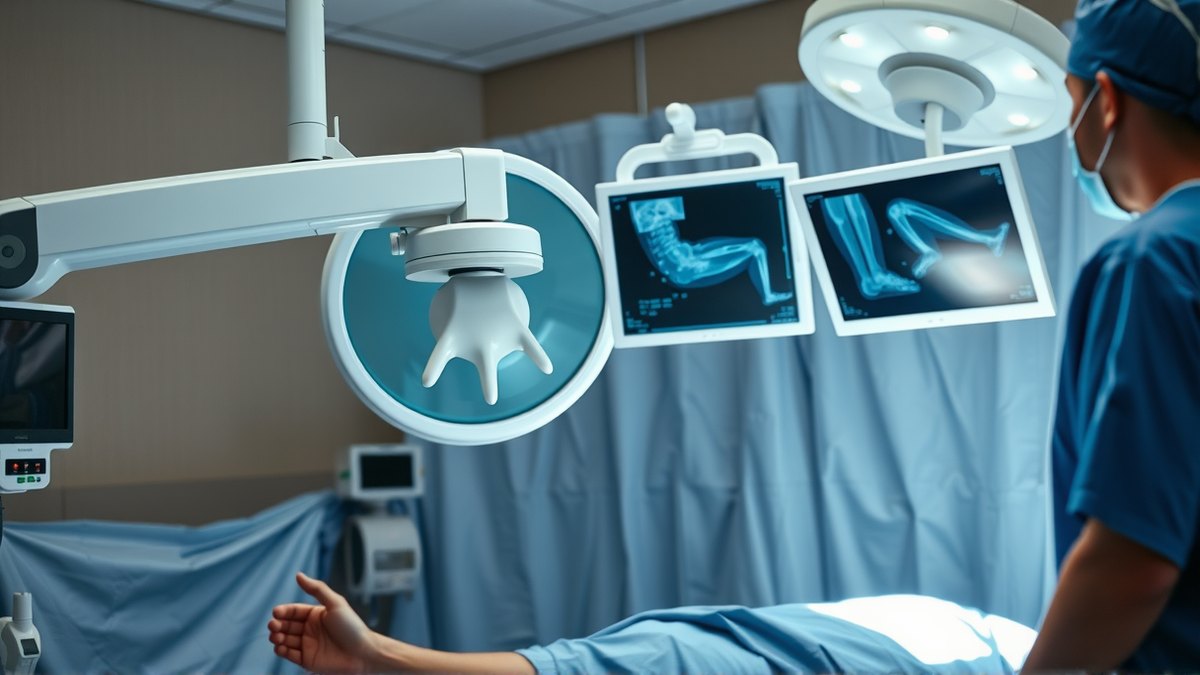
In minimally invasive procedures such as angioplasty, the doctor guides instruments through a small incision and watches their position on a live X-ray image. An X-ray image carries only two dimensions of information. It is hard to tell exactly where the instrument sits inside the patient's body and how it is oriented. Matching an X-ray by hand to a preoperative CT or MRI takes time, and existing AI tools handled it inconsistently from one patient to the next.
A team at the Massachusetts Institute of Technology and collaborating institutions built a system called xvr. It does not try to train one model for all patients. Instead it adapts to a specific person. From that patient's preoperative CT or MRI data it generates simulated X-ray images from various angles, reproducing the physical processes that form the image. The system produces about 1000 simulated images per second, and the simulation draws only on the patient's own data. It is not generating data out of nothing, so there is no hallucination problem.
From twelve hours to five minutes
Training a model dedicated to a single patient from scratch would take about 12 hours. The teams therefore used 3D scans of more than 2000 patients to pretrain a base model. For a new person, fine-tuning then takes about five minutes, after which the matching itself finishes in a few seconds.
In tests on real data from five hospitals, the system covered adult and pediatric patients and several dozen bone and organ setups. xvr proved more accurate and more stable than existing AI methods, and at the same time fast enough for emergency settings, for example in an operating room. The results were published in the journal Nature.
The practical value is twofold. Faster and more reliable image registration helps the doctor judge the position of a catheter or endoscope and lowers the risk of damaging surrounding tissue. The same technology can serve to navigate surgical robots.
Uncertainty has to be measured too
Image registration is not the only front in work on trust in imaging models. In the list of medical physics preprints on arXiv, published continuously, work appeared in the same week on artifact reduction in cone-beam imaging (arXiv:2609.30169), on a generative substitute for Monte Carlo calculations in photoacoustic imaging (arXiv:2609.28261), and on identifiable uncertainty in CT imaging (arXiv:2609.29599). The last of these matters for clinical use: a model that can say how unsure it is of its own result is easier to plug into a decision process.
Sources
2All figures and quotations in this text come from the sources listed below.
Content prepared by the editorial team with AI assistance.
Sofia Marchetti
Comments
0- No comments yet — be the first.