One spine MRI scan, 76 diseases, and a draft report ready to check
The radiology department at Peking University Third Hospital and United Imaging Intelligence built an agent that turns a routine spine MRI into a structured report covering 76 diseases. Report writing went 56 percent faster.
 Sofia MarchettiScience and health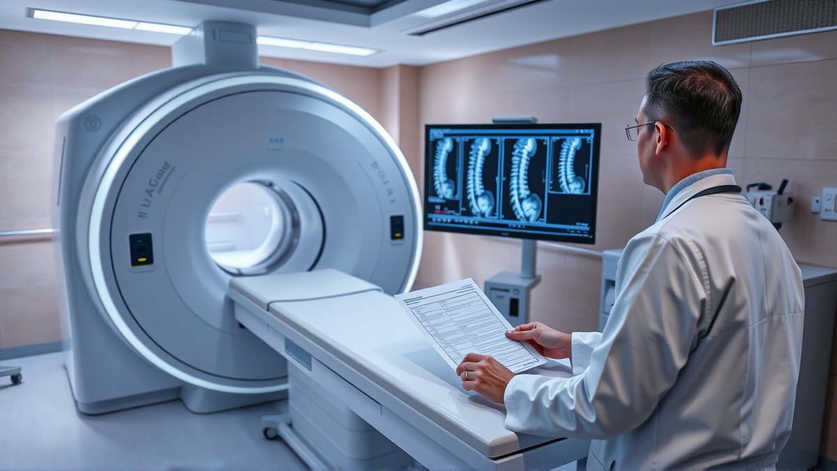
Medical imaging is full of AI models that shine on laboratory data and then stumble in a real clinic. The question is no longer whether the technology works. It is how to get from technical availability to clinical usefulness.
The radiology department at Peking University Third Hospital and United Imaging Intelligence built a spine MR agent that works on a one scan, many examinations principle. A single spine MRI yields a structured analysis of several conditions at once. Prof. Yuan Huishu, who heads diagnostics at the department, says the agent combines a large model with the way clinicians actually work, so the information is complete, precise and easy to use. It takes repetitive observation, sorting and typing off the doctor's hands.
Scale of the burden
Spine MRI is one of the most common examinations in the imaging suite. At this Beijing hospital it is the single most common MR examination in the whole facility, averaging close to 300 scans a day. Reading one means going layer by layer through vertebrae, discs, ligaments, spinal cord and nerve roots, then describing herniations, canal stenoses or changes in the vertebral bodies. Without AI support, a report takes five to ten minutes on average from first draft to approval.
Prof. Yuan points out that traditional AI models solve one task or a few. They act like specialist observers of a single type of lesion. They are also often separate programs outside the PACS system, so the doctor has to move results and order the text by hand. That adds work instead of removing it. Some tools produced answers that did not match the image, and that undermined trust in the technology itself.
Three thresholds
The agent makes three jumps. First, from spotting a single disease to assessing many at once. It reads the image globally and produces a report covering 76 diseases and abnormalities in one pass, going through the spine sections one by one. In a lumbar examination for back pain it picks up not only disc degeneration but also vertebral haemangiomas or compression fractures. Second, from marking foci to a full structured report: detection of a lesion, anatomical localisation, description of findings and a ready structured report all come out together. Report writing efficiency rose by 56 percent. Third, from an external tool to an agent embedded in the AI PACS platform the doctor uses every day.
The project formally began in July 2025. It followed an 18-month overhaul of the AI PACS platform carried out by both sides. The team lists four technical challenges: precise anatomical localisation of the many spine sections, understanding the logic radiologists use to write reports, the long tail of rare diseases with too little data, and keeping the model from hallucinating. The agent already works at the main hospital and four branches. AI remains an auxiliary tool: a radiologist must approve every result. The next stage is an external multicentre study.
Sources
2All figures and quotations in this text come from the sources listed below.
Content prepared by the editorial team with AI assistance.
Sofia Marchetti
Comments
0- No comments yet — be the first.